Medneo accelerates growth and enters veterinary imaging with acquisition of Burgess Diagnostics
21 Sep 2026
We are pleased to announce that Medneo has acquired Burgess Diagnostics Ltd, 21st September 2026. Read more
Middle-aged Labrador Retriever.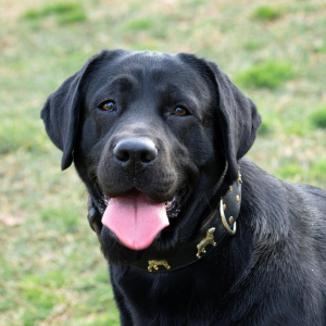
The patient was fit and active but presented with three, short, grand-mal seizures which responded to levetiracetam. Their general physical, orthopaedic, and neurological exam was normal.
Haematology, biochemistry, ionised calcium, glucose, and bile acid stimulation test all normal. Platelet count and coags – normal.
Number of series / images: 12 / 349 Series: Transverse plane T2W, FLAIR, T2*GRE, T1W pre and post-contrast, DWI with ADC map. Sagittal T2W. Dorsal T1W pre and post-contrast.
Figure 1: Left to right: parasagittal T2W, transverse plane T2W, dorsal plane T1W precontrast, dorsal plane T1W post-contrast.
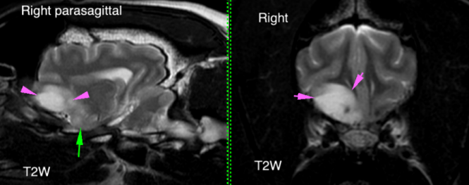
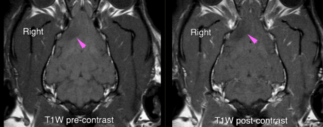
Figure 2: Diffusion-weighted image series
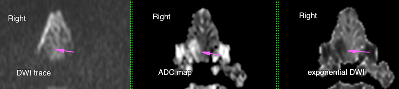
A soft tissue mass is present in the right rostroventral part of the cerebrum, in the region of the right frontal lobe, centred on cerebral grey matter (Figure 1, Pink arrows). Affected grey matter is swollen and T2W hyperintense, mildly hyperintense on FLAIR and T2*, hypointense on T1W and does not contrast enhance. There is NOT abnormal restricted diffusion, with the lesion hypointense on trace and exponential DWI and hyperintense on ADC map representing T2W shinethrough (Figure 2). There is poorly defined T2W and FLAIR hyperintensity within the right temporal cortical grey-matter immediately caudal to the mass (Figure 1, left, green arrow). White matter is spared. The mass causes moderate mass effect with displacement of the falx cerebri to the left and compression and displacement of the rostral horn of the right lateral ventricle.
Right frontal cortical mass. ddx: neoplasia such as a nonenhancing glioma, glioblastoma, less likely diffusion negative acute ischemic infarct (rare and unlikely given 6 week duration of signs).
CSF – normal.
CT thorax and abdomen – declined for financial and ethical reasons (we would be searching for the primary tumours in a dog with metastatic disease).
Surgery (biopsy, or attempted excision using neuronavigational aids and intra-operative photodynamic detection (with eg 5-ALA (“The Pink Drink”)), plus radiotherapy). Chemotherapy (temozolomide) – not reported as a sole-agent in the face of gross disease, only as an adjunct for surgery/radiotherapy. Palliative - 1000mg levetiracetam TID to control signs, consider adding 0.5-1mg/kg prednisolone if the signs progress, or phenobarbitone 3mg/kg BID if seizure frequency increases.
Intracranial gliomas in dogs are associated with a poor prognosis, however a combination of surgery, chemotherapy, and radiotherapy can increase the survival time from around 1-3 months with palliative care, to up to 1.5 years with radiotherapy or surgery plus radiotherapy +/- chemotherapy.
Surgery, radiotherapy, and chemotherapy were declined in this case for ethical, practical, and financial reasons.
Gliomas are not commonly seen in Labrador retrievers so we do need to keep in mind benign differentials in mind eg granulomatous lesion. In the event the clinical picture is stable in three months’ time, repeat MRI is recommended. If the lesion is shrinking the prognosis would be better.
We are always tempted to give steroids but at this stage there is no perilesional oedema that would benefit from steroids so we have not recommended steroids for now.
The patient had an extension to her good quality and length of life, on treatment with levetiracetam. Five months after diagnosis, when her signs progressed despite treatment, the owners were able to make the final decision for her to prevent any suffering. Because of the MRI findings and her diagnosis, her owners had full confidence that they had done everything they could, and could put her best interests at the heart of their decision. Rest in peace.
Ian Nicholson BVSc CertSAS DipECVS MRCVS RCVS and ECVS Specialist in Small Animal Surgery, Island Referrals

21 Sep 2026
We are pleased to announce that Medneo has acquired Burgess Diagnostics Ltd, 21st September 2026. Read more
14 Sep 2026
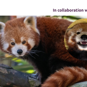
In August 2026 the Burgess Diagnostics mobile imaging team had the privilege of supporting the Animal Care team at Colchester Zoo for a full day of advanced CT imaging. The visit formed part of Colchester Zoological Society’s commitment to delivering exceptional standards of animal care, using this state-of-the-art diagnostic tool to better understand the health of the extraordinary animals in their care. Read more