New CT Referrals - North Wales
6 Jul 2026
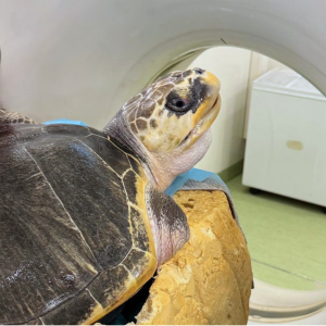
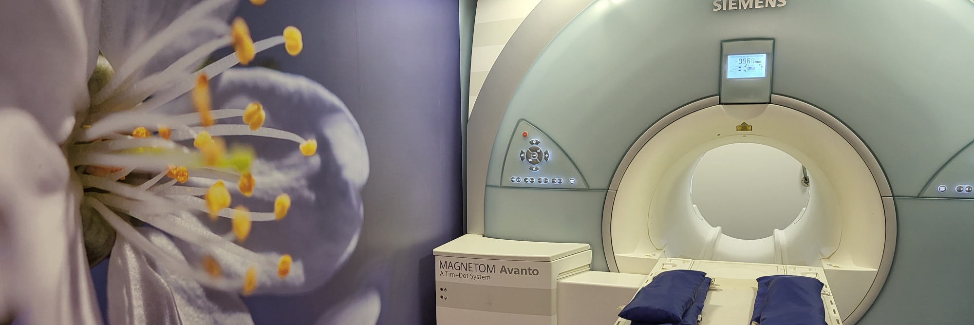
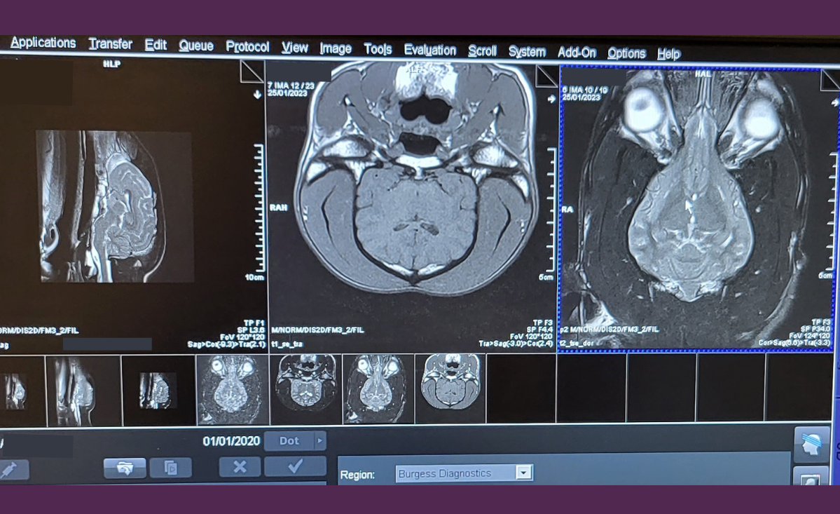
The fabulous team at North Wales Referrals will now offer CT imaging alongside their core referral services, which include soft tissue and orthopaedic surgery, cardiology, internal medicine, physiotherapy, hydrotherapy, and laser therapy. Read more