Linkedin Email us 0845 3714 012

MR imaging of seizures in a dog

An adult Bichon Frise first presented, aged 9 years, with seizures. CSF, bloods and MRI at initial presentation were all said to be normal and the dog responded well to medical treatment.

Seizures 1

Initial MRI (T2W TSE)

Some nine months later, deterioration in seizure control occurred with the dog experiencing a cluster of seizures. Medication was increased and the patient was referred for a second MRI. On this occasion an ill-defined area of T2W hyperintensity was found in the ventral aspect of the right temporal lobe and hippocampus with corresponding T1W hypointensity.

Seizures 3Seizures 2 

T2W TSE                                                                                                                                       T1W IR

Diffusion weighted imaging was not available at the time of the initial scan as this was carried out on a low-field scanner but DWI on this occasion demonstrated no evidence of restricted diffusion. A diagnosis of non-haemorrhagic infarct was made.

Seizures 4

DWI


Latest News View all news


PHOTO-2025-10-27-14-10-59

Groundbreaking CT Scans Deliver Vital Answers!

6 May 2026

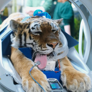
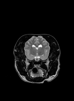
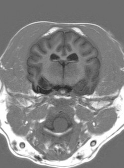
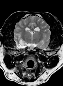
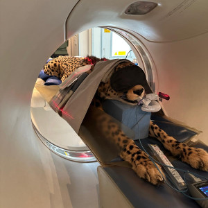
Discover how the ground breaking CT scans we performed at The Big Cat Sanctuary in October have helped their team to refine treatment plans, make informed decisions, and safeguard each cat’s long-term welfare. Read more

width_533

The tiger who came for CT

21 Apr 2026

Advanced CT Imaging Supporting Wildlife Care and Conservation at Banham Zoo - Find our more about our CT scanning day at Banham Zoo where we scanned a tiger, a sloth, a vulture and many more exotic species. Read more

Burgess Diagnostics uses cookies to ensure that we give you the best experience on our website. Please accept cookies for optimal performance. For more information please visit our cookies policy.