NEW CT SERVICE LAUNCHES AT LANES VETS!
11 Mar 2025
The Burgess Diagnostics mobile CT service will now be visiting Lanes Vets in Garstang, to assist them with their cases. Read more
The prevalence of primary prostatic cancer in dogs is low (0.6% at necropsy), occurring more frequently in older dogs with a mean age of 9.9 years.
Ca Prostate is more common in certain breeds with Bouvier des Flandres, Doberman Pinschers, Scottish Terriers and Shetland Collies some of the breeds particularly affected.
Most primary prostate cancers are aggressive and highly metastatic, the most prevalent type being prostatic adenocarcinoma. Tumour growth outside the prostate is common, as are pulmonary and bone metastases. Other symptoms witnessed may include:
Stranguria, Haematuria, Pollakiuria, Colon compression causing difficulty in defaecation.
Dogs with metastatic spread are more likely to present with primarily neurological symptoms such as:
Lameness, Ataxia, Paresis, Paralysis
Lung metastases are found to be present in 44% of cases at diagnosis.
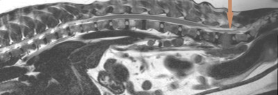
A 7-Year old Scottish Terrier was referred for MRI imaging of the thoraco-lumbar spine. The clinical presentation was of acute onset of hindlimb lameness over the preceding 3-4 weeks with hyporexia and lethargy. Localised pain was noted in the L6 region on clinical examination.
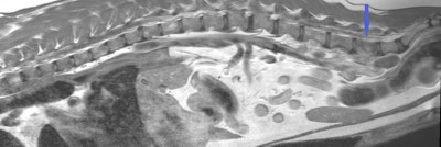
Initial sagittal imaging of the thoraco-lumbar spine revealed infiltrative lesions in the L5 and L6 vertebral bodies. These lesions were evidenced by marked cortical lysis of the ventral aspect of the vertebral bodies and displayed ill-defined T1 hypointensity when compared to normal bone marrow, and heterogenous mixed T2 intensity.
T2 Sagittal T/L Spine (L6 Lesion – Brown Arrow)
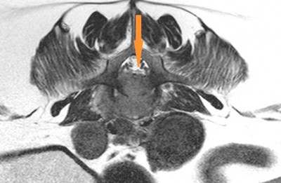
T2 Transverse through L6
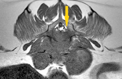
T1 Transverse through L6
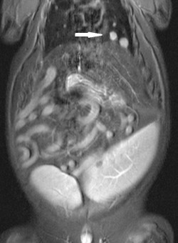
Dorsal STIR imaging throughout the T/L Spine, abdomen and lower half of the thorax revealed the presence of multiple rounded hyperintense lesions spread throughout the visible lung lobes, varying in size from 3 – 13mm. These lesions were indicative of metastatic deposits (White Arrow below)
Dorsal STIR
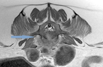
Post Contrast imaging of the spinal lesions displayed moderate heterogenous contrast enhancement of the infiltrated vertebral bodies (Blue Arrow), allied to irregular contrast enhancing periosteal reaction. Additionally, a mildly enhancing, heterogenous soft-tissue lesion extending from the ventral aspect of was L6 evidenced, extending ventrally and predominantly on the right side into the paraspinal musculature (Teal Arrow).
T1 Sagittal Post Contrast
T1 Transverse Post Contrast
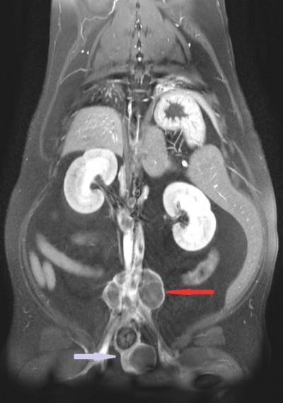
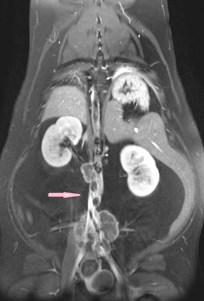
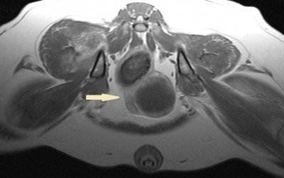
Dorsal post-contrast T1 Fat Saturation imaging revealed extensive enlargement of the para-aortic and internal iliac lymph nodes, with a heterogenous rim-enhancement (Pink / Red Arrows). Additionally, the prostate gland is markedly enlarged with capsular thickening on the right side and heterogenous contrast enhancement, most noticeably on the rim (Purple Arrow).
DORSAL T1 FAT SAT IMAGES
The dorsal imaging sequences also revealed marked cortical osteolysis of the left proximal femur with associated contrast enhancement of the bone and soft tissues on the left, with ill-defined contrast enhancement on the contra-lateral side (Yellow arrow below).
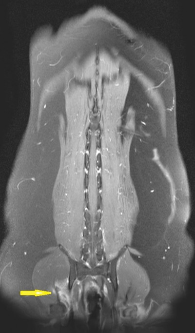
T1 transverse post-contrast imaging through the pelvis better illustrates the cystic nature of the prostate mass, as well as the degree of wall thickening previously described (Cream Arrow).
PROBABLE DIAGNOSIS
Marked aggressive prostate mass most likely consistent with adenocarcinoma. Extension of the neoplastic process in the surrounding soft tissue, paravertebral soft tissue, retroperitoneum and regional metastasis to the sub-lumbar and aortic lymph nodes, with osseous metastasis to multiple sites. Abundant distant pulmonary metastases are also noted.

11 Mar 2025
The Burgess Diagnostics mobile CT service will now be visiting Lanes Vets in Garstang, to assist them with their cases. Read more

10 Dec 2024
We are pleased to announce that the Burgess Diagnostics mobile MRI service made its first-ever visit to South East Vet Referrals, in Kent, today. Read more