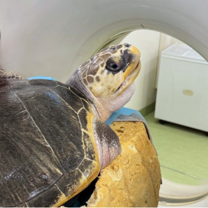
Linkedin Email us 0845 3714 012

MR imaging of seizures in a dog

An adult Bichon Frise first presented, aged 9 years, with seizures. CSF, bloods and MRI at initial presentation were all said to be normal and the dog responded well to medical treatment.

Seizures 1

Initial MRI (T2W TSE)

Some nine months later, deterioration in seizure control occurred with the dog experiencing a cluster of seizures. Medication was increased and the patient was referred for a second MRI. On this occasion an ill-defined area of T2W hyperintensity was found in the ventral aspect of the right temporal lobe and hippocampus with corresponding T1W hypointensity.

Seizures 3Seizures 2 

T2W TSE                                                                                                                                       T1W IR

Diffusion weighted imaging was not available at the time of the initial scan as this was carried out on a low-field scanner but DWI on this occasion demonstrated no evidence of restricted diffusion. A diagnosis of non-haemorrhagic infarct was made.

Seizures 4

DWI


Latest News View all news


Untitled design (24)

Meeting Kiburi - the Gorilla!

22 Sep 2025

It’s not every day we welcome a gorilla on board one of our mobile CT scanners! Find out more about our amazing day at London Zoo. Read more

japanese_macaque_1

A wild scanning day at the Highland Wildlife Park

1 Sep 2025

From macaque's to meercats, a scanning day to remember. Find out more about our incredible day at the Highland Wildlife Park. Read more

Burgess Diagnostics uses cookies to ensure that we give you the best experience on our website. Please accept cookies for optimal performance. For more information please visit our cookies policy.