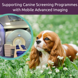
Magnetic Resonance Imaging
What is Magnetic Resonance Imaging (MRI)?
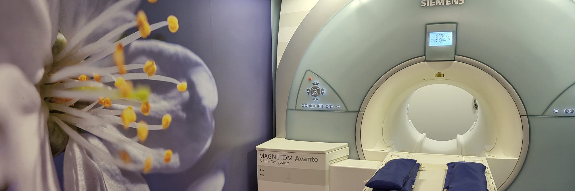
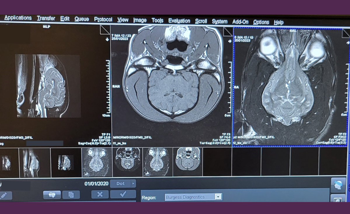
Magnetic Resonance Imaging (MRI) is based on the use of strong magnetic fields and radiofrequency pulses to generate cross-sectional images. Different combinations of these are used to create sequences of images with different contrast and many different sequences are available. This, combined with imaging in any plane (sagittal, dorsal, transverse, oblique) and without the associated ionising radiation (which is used in CT) makes MRI the optimum method of investigation in the majority of clinical cases. It is unparalleled in the investigation of soft tissues due to its superior contrast sensitivity and tissue discrimination.
For accurate diagnosis and lesion localisation, MRI is now the investigation of choice in all veterinary neurological, joint, and spinal disease processes. With multi-planar capabilities, various MRI sequences, and the latest contrast agents, tissue and disease characterisation will allow accurate identification, case treatment, and management.
What are the key advantages of Magnetic Resonance Imaging (MRI)?
- Images available in any plane
- Clear anatomical images of the highest quality and clarity
- Greater soft tissue contrast (compared to CT)
- Uses non-ionising radiation
- Non-invasive procedure
- Numerous advanced techniques can be performed with high-field scanners
- An excellent diagnostic tool with a wide range of clinical applications
- Safe and cost-effective
What are the clinical applications of Magnetic Resonance Imaging (MRI)?
- Spinal disorders (e.g. Diskospondylitis, Intervertebral Disc Disease, Neoplasia)
- Brain conditions (e.g. Intracranial Vascular Disease, Encephalitis, Haemorrhage, Oedema, Epilepsy, Metastasis)
- Nasal, oral & optic conditions (e.g Osteomyelitis , Aspergillosis, Palatine Destruction)
- Ear conditions (e.g. Tympanic Bulla Wall Erosion, Neoplasia, Lymphadenopathy)
- Joint problems (e.g. Bone Enema, Osteochondritis, Arthrograms, Muscular Injuries, Cruciate Ligaments)
- Abdomen & pelvic regions (e.g Prostate, Hips, Ilio-psoas Injury)
- Thorax (e.g. Thoracic Wall Mass, Mediastinal Mass)
For guidance on choosing the optimum imaging modality when considering specific anatomical regions please click here