
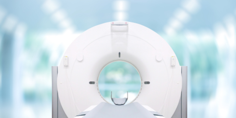
What is Computed Tomography (CT)?
Computed Tomography (CT), commonly referred to as a CAT scan, is a cross-sectional imaging modality that uses x-rays to produce a cross-sectional, or ‘slice’ image of the inside of the body. The process can show bones, as well as surrounding soft tissues such as muscle and blood vessels with excellent clarity. CT imaging produces a high volume of data which can be manipulated electronically to allow reformatting in multiple planes.
Current multidetector CT (MDCT) technology allows rapid imaging and generates a volume of attenuation data which enables multiplanar and 3D reconstructions. MDCT also facilitates highly detailed CT angiography using iodinated contrast media.
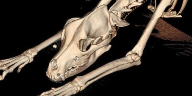
The Burgess CT systems can quickly scan large areas of the patient, in one continuous operation, and deliver large numbers of images to help in the diagnosis of many conditions in small animals. These can include neoplasm, infectious disease, trauma, and musculoskeletal disorders.
What are the key advantages of Computed Tomography (CT)?
- Minimal anaesthetic/sedation time required for the patient
- Produces images of the highest quality and clarity
- Images acquired in the Axial plane and can be reconstructed in the sagittal and dorsal planes
- An excellent diagnostic tool with a wide range of applications
- CT angiography is easy to perform
What are the clinical applications of Computed Tomography?
- Head/Neck – Nasal disease, orbital swelling, head trauma, ear disease, thyroid masses, neck swellings
- Chest – Pleuritis, rib masses, sternal/mediastinal masses, lung tumours, lymph nodes, metastatic screening, pneumothorax
- Abdomen – Liver and/or abdominal masses, ectopic ureters, pancreatitis, insulinoma, adrenal masses, portosystemic shunts, retroperitoneal effusion or masses
- Spine & Pelvis - Vertebral anomalies and malformation, lumbosacral disease, pelvic and sacral fractures, bone tumours
- Orthopaedic - Tarsal osteochondritis, bone tumours, elbow dysplasia, angular limb deformity
For guidance on choosing the optimum imaging modality when considering specific anatomical regions please click here.

