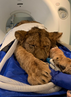
CT imaging of a Lioness
Computed tomography, (CT) is a very affective diagnostic tool used widely in veterinary medicine for detailed diagnostics images and tends to be the imaging of choice for many vets, due to its speed, versatility and accuracy.
Patient:
- Panthera Leo (Lion/Lioness)
- 10 months old
- 78 Kg
Clinical details:
- Presented with acute 3-4 days of blindness
- Ataxia (lack of muscle control)
- Elevated bile acids
- Off colour
- Elevated liver enzymes
Clinical question:
- Liver pathology (shunt)
- Neurological lesions
- Abdominal abnormality
Clinical plan:
- CT head pre/post contrast
- CT Chest
- CT Abdomen & Pelvis
CT scans:
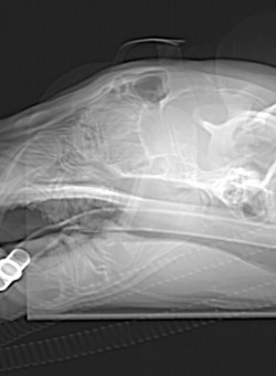
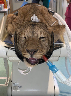
The brain was the first thing to image as position would have to change for the abdominal study. Scan acquisition was from tip of nose down to C2. A lateral and AP survey was taken to ensure all anatomy is included in the scan. The recons are just as important and a bony 0.6mm recon is required to successfully assess all structures.
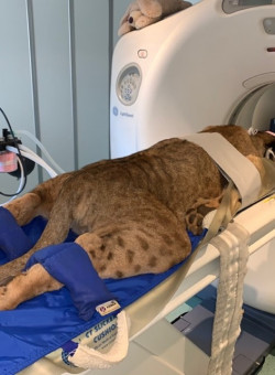
For the abdominal scan we turned the patient and positioned her prone, which is important for assessing the liver and additional structures within the abdominal cavity.
The abdominal scanning consisted of three phases in quick concession. A non-contrast, arterial and a portal venous phase, if the contrast has not been visualised correctly another delayed scan should be performed. Anatomy wise, acquisitions should start from above the diaphragm to past the anal glands of the patient too ensure all structures visualised and can be assessed.
Diagnosis
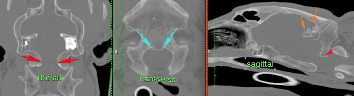
The brain CT report stated that, ‘irregular thickening of the occipital bones, resulting in mild crowding of the caudal cranial fossa and narrowing of the foramen magnum which results in mild compression of the brain stem’, which is called calvarial hyperostosis.
The crowding within the caudal brain stem explains the neurological signs the patient was symptomatic, with blindness which occurs within hypovitaminosis A, (suspected diagnosis).
Results
Within the abdomen it was reported that the liver showed a suspicion of multiple small aberrant vessels at the level of the coeliac artery. Interestingly, Vitamin A is stored within the liver so the hepatic pathology may have changed the concentrations within the liver leading to hypovitaminosis A, equally added to poor retinol and dietary intake. The changes within the skull, and the liver appearing relatively normal with no shunt visible. It was concluded that this could be related to hypovitaminosis A, this is a lack of vitamin A within the blood and tissue of the patient.
Using CT imaging in multiphase scanning diagnosed the Panthera Leo and a treatment plan was undertook and is recovering well.